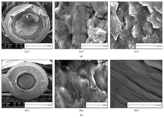
Figure 3.
Failure analysis of retrieved fractured Ti-6Al-4V implant (a) and CP-Ti implant (b). (A1) and (B1) are macroscopic views of the fracture surface of the implant. (A2) and (B2) show fatigue striations on retrieved fractured implants. (A3) and (B3) show fatigue striations on dental implants fractured in laboratory conditions in room air. Note the high resemblance of the in vivo and in vitro fracture surface topographies.

