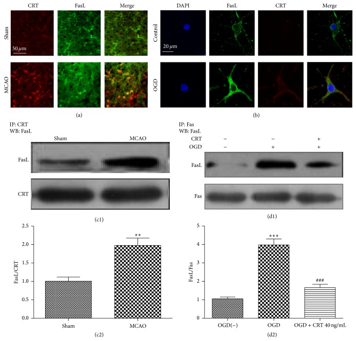
Figure 2.
CRT binds to FasL on the surface of neurons after ischemia. (a) Immunofluorescent staining of the cortexes of sham and MCAO mice. (b) Confocal microscopy images of immunostained normal SH-SY5Y cells and OGD-exposed SH-SY5Y cells at 3 h after reperfusion. (c1, 2) Co-IP of CRT and FasL in the cortexes of sham and MCAO mice. (d1, 2) Co-IP of Fas and FasL in normal SH-SY5Y cells and OGD-exposed SH-SY5Y cells cultured with or without 40 ng/mL CRT. ∗∗ P < 0.01, ∗∗∗ P < 0.0001 versus control, and ### P < 0.0001 versus OGD group. n = 6 repeats.

