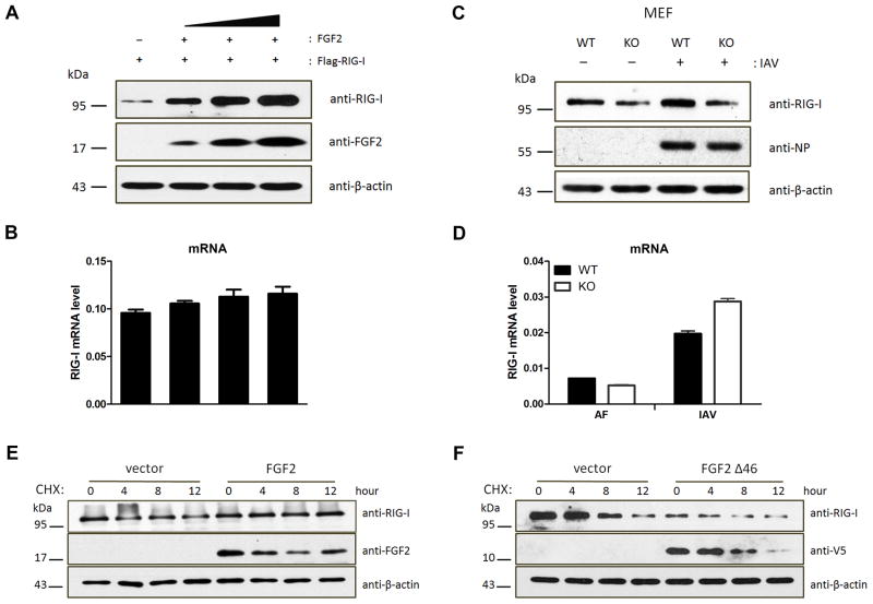
Figure 5. LMW FGF2 increases RIG-I stability.
(A, B) 293T cells were transfected with Flag-RIG-I and various amounts of LMW FGF2 (20, 50, and 100 ng/μl). Forty-eight hours after transfection, (A) WCLs of 293T cells were subjected to an IB assay using anti-RIG-I, anti-FGF2, and anti-β-actin antibodies; (B) The RIG-I mRNA levels were assessed by real time-PCR.
(C, D) Primary MEFs isolated from Fgf2lmw+/+ (WT) or Fgf2lmw−/− (KO) mice were infected with IAV or allantoic fluid (AF). (C) Cell lysates were harvested at 12 hpi and then subjected to IB assay using anti-RIG-I, anti-influenza A virus nuclear protein (NP) and anti-β-actin antibodies; (D) The RIG-I mRNA levels were assessed by real time-PCR.
(E, F) 293T cells were transfected with plasmid encoding LMW FGF2 (E), LMW FGF2 Δ46 (F) or empty plasmid as control. Forty-eight hours later, 293T cells were treated with 100 μM cycloheximide (CHX). The cells were harvested at the indicated times and then subjected to an IB assay using anti-RIG-I, anti-FGF2 or anti-V5 and anti-β-actin antibodies.

