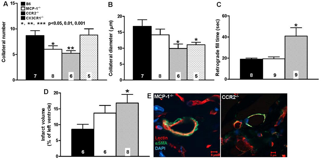
Figure 5. Neo-collateral formation 1 week after LAD ligation is dependent on MCP-1 →CCR2 signaling.
A–D, number, diameter, retrograde fill time and infarct volume determined as in Figure 3. CCR2−/− mice have impaired neo-collateral formation. “conductance” and increased infarct volume. MCP-1−/− show smaller difference from C57BL/6 (B6), possibly owing to other MCP isoforms acting at CCR2. E, Neo-collaterals of both MCP-1−/− and CCR2−/− mice have αSMA+ mural cells; magnification bars, 5 um; data are representative of ≥ 3 mice of each. Statistics are versus B6 (background of knockout mice).

