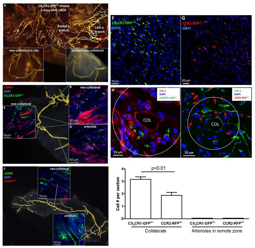
Figure 6. CCR2RFP/+ and CX3CR1GFP/+ B6 reporter mice show both cell types present throughout border zone and in vicinity of developing neo-collaterals 5 days after LAD ligation.
A–E, Whole mount images. αSMA+ mural cells surround neo-collaterals and myofibroblast-like cells present in interstitium. Almost no CCR2+ or CX3CR1+ cells seen in remote zone and none within the vicinity of arterioles or venules (Online Figure 17). F,G, 7µm sections from border zone; many fewer CCR2+ or CX1CR3+ cells seen 1 day after LADX (n=5 mice, data not shown). H, Neo-collaterals (COL) in border zone. Upper panels: arrows identify both cells types; Flk1+ cells identify endothelial cells of collateral and nearby vessels, and possible monocyte-like EPCs (*). Quantification: cell number counted within area extending 1-collateral diameter outward and averaged for 5 sections separated by 50µm from midpoint of 1 neo-collateral for each mouse; 5 mice 5 days after LADX were studied.

