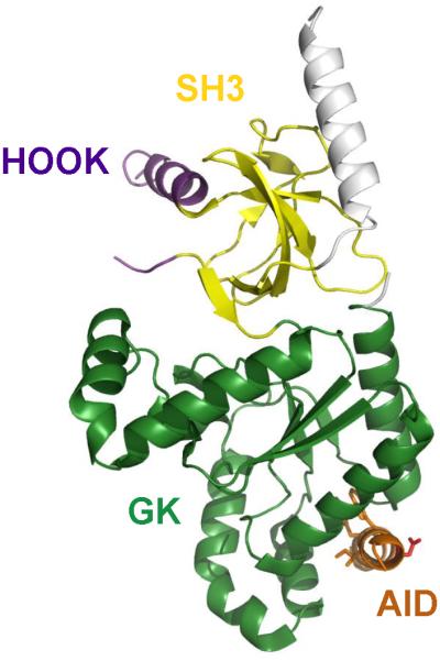
Figure 9. Structural model of the CaVβ subunit.

The SH3 domain (yellow), HOOK domain (purple), Alpha Interaction Domain (AID, red), and Guanylate Kinase domain (GK, green) are illustrated in the indicated colors.

The SH3 domain (yellow), HOOK domain (purple), Alpha Interaction Domain (AID, red), and Guanylate Kinase domain (GK, green) are illustrated in the indicated colors.