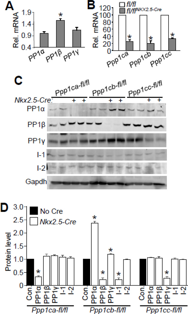
Figure 1. Generation and assessment of cardiac specific PP1 isoform deleted mice.
(A) Real-time PCR analysis of expression of PP1 isoforms in adult cardiac myocytes. Rpl27 was used as an internal control. N=3 for each group. *p<0.05 vs PP1α. (B) Realtime PCR analysis of expression of PP1 isoforms in the hearts of 2 month-old mice for each of the groups shown, with or without Cre-mediated deletion. N=4 for each group in the real-time PCR analysis. *p<0.05 vs fl/fl mice. (C) Western blots for PP1 isoforms, I-1, I-2, and gapdh from the hearts of Ppp1c-fl/fl mice or Ppp1c-fl/flNkx2.5-Cre mice. Approximately 120 µg of protein was processed to detect I-1 and I-2. D) Quantification of Western blots shown in Figure 1C. N=4 for each of the groups. *p<0.05 vs control (Con.).

