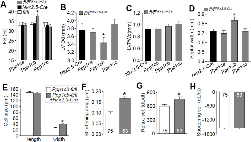
Figure 2. Cardiac deletion of PP1β leads to enhanced contraction and cardiac remodeling.
(A–D) Echocardiographic measurements of 2 month-old mice for fractional shortening (FS%), left ventricular internal diastolic dimension (LVIDd), left ventricular posterior wall dimension(LVPWd), and septal width. N=10 for each group. *p<0.05 vs Nkx2.5-Cre. (E) Length and width of 224 adult cardiac myocytes isolated from 2 month-old mice of the indicated genotypes, measured by NIH ImageJ software.*p< 0.05 vs Ppp1cb-fl/fl.(F–H) Shortening amplitude, relaxation velocity, and shortening velocity in isolated adult cardiomyocyte from the hearts of the indicated genotypes of mice. *p<0.05 vs Ppp1cb-fl/fl. Data are normalized to the peak of their shortening amplitude, and units are µm/second for change in length (dL) per change in time (dt).

