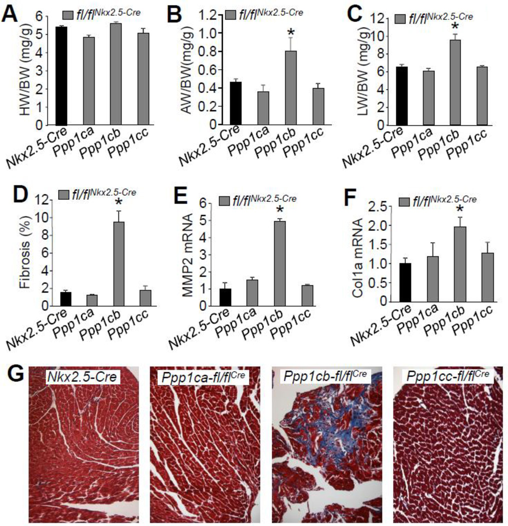
Figure 3. Cardiac deletion of PP1β results in interstitial fibrosis and heart failure.
(A–C) Measurements of heart weight/body weight (HW/BW), atria weight/body weight (AW/BW), and lung weight/body weight (LW/BW) in the indicated groups of mice, assayed at 2 months of age. N=8 for each group of mice. (D) Percentage of interstitial fibrosis from the hearts of the indicated mice, as quantified using MetaMorph software based on Masson’s trichrome-stained heart histological sections as shown in panel G. Images from at least 4 hearts in each group were used for analysis. (E–F) Quantitative PCR analysis of expression of MMP2 and collagen 1 α1 using total RNAs purifed from hearts of each of the groups indicated (N=3). (F) Representative histological sections stained with Masson’s trichrome (shows fibrosis in blue) from the hearts of the indicated genotypes of mice. *p<0.05 vs Nkx2.5-Cre for all panels.

