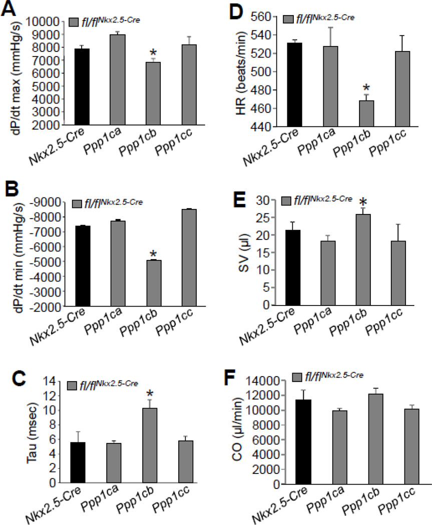
Figure 4. Cardiac deletion of PP1β protein results in contractile dysfunction.
(A–B) Invasive hemodynamic measurement of cardiac contractility assessed at baseline (load-dependent) as maximum and minimum rate of pressure change in the left ventricle over time (dP/dt max and dP/dt min, respectively) in Nkx2.5-Cre control mice versus Ppp1ca-fl/flNkx2.5-Cre, Ppp1cb-fl/flNkx2.5-Cre, and Ppp1cc- fl/flNkx2.5-Cre containing mice (C) Time constant for isovolumetric relaxation (Tau) showing the exponential decay of ventricular pressure during isovolumic relaxation. (D–F) Analysis of heart rate (HR), stroke volume (SV), and cardiac output (CO) from the hemodynamic measurements in the indicated genotypes of mice. At least 3 mice for each group were used for the measurements. *p<0.05 vs Nkx2.5-Cre.

