
Fig. 2. Increased phospho-ERK1/2 staining in the rat brain in KA-SE.
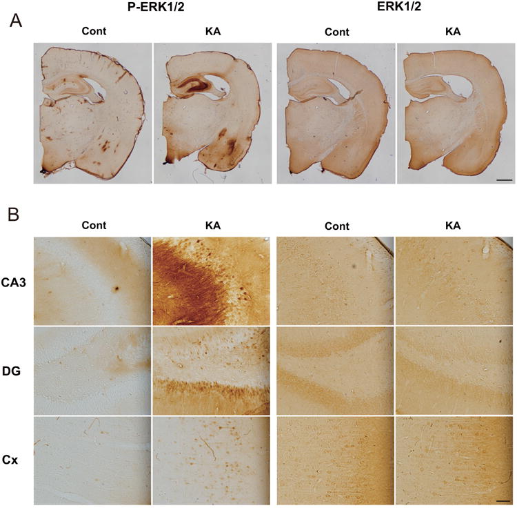
A, Representative staining with anti-phospho-ERK1/2 (P-ERK1/2) and anti-ERK1/2 antibodies in a frontal section. Intense phospho-ERK1/2 staining was observed in the hippocampus, amygdala and piriform cortex from rats in KA-SE (KA), but not in control (Cont). Total ERK1/2 staining was unchanged between KA-SE and control. B, Phospho-ERK1/2 and total ERK1/2 staining with higher magnification in the CA3 and dentate gyrus (DG) of the hippocampus, and the lateral portion of the cerebral cortex (Cx), corresponding to the parietal cortex. Phospho-ERK1/2 staining was especially increased in the stratum radiatum of the CA3 region and in the granular cell layer of the dentate gyrus. Total ERK1/2 staining was similar between KA-SE and control. Scale bars, 1000 μm in A, 50 μm in B.