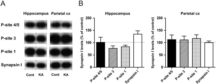
Fig. 4. Recovery of phospho-synapsin I levels in brain homogenates from rats in KA-recovery.

A, Representative immunoblots in the hippocampus and parietal cortex (Parietal cx) from rats in KA-recovery (KA), as compared to those from controls (Cont). Decreased phospho-site levels observed in KA-SE (Fig. 3) were no longer detected in KA-recovery. B, Quantitative data obtained by immunoblot analyses, expressed as a percentage of control levels (n=5). No significant changes were observed in any phospho-site levels, as well as in the total synapsin I level, in both brain regions.
