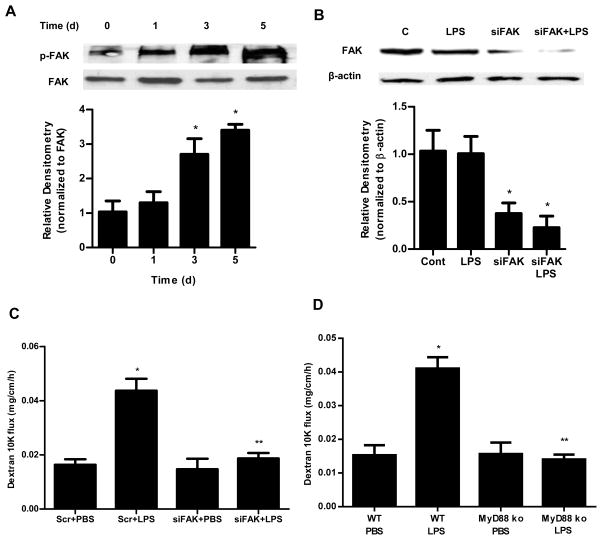
Figure 7.
Involvement of FAK and MyD88 in LPS-induced increase in mouse intestinal permeability. (A). Time-course effect of LPS (0.1 mg/kg body weight) on phospho-FAK in mouse intestinal tissue as assessed by Western blot analysis. Densitometry analysis showed significant time-dependent increase in the expression of phospho-FAK. n=3. (B). FAK siRNA silencing knocked down the expression of FAK in mouse intestinal tissue as assessed by Western blot and densitometry analysis. siFAK transfection was performed 1 d prior to the LPS treatment, and the Western blot analysis was performed after 5 d LPS treatment. n=3. (C). Effect of siRNA-induced silencing of FAK on LPS-induced increase in mouse intestinal permeability. n=4. (D). Effect of LPS on intestinal permeability in MyD88−/− mice. n=4. *, p < 0.001 vs WT control; **, p < 0.001 vs WT LPS treatment.

