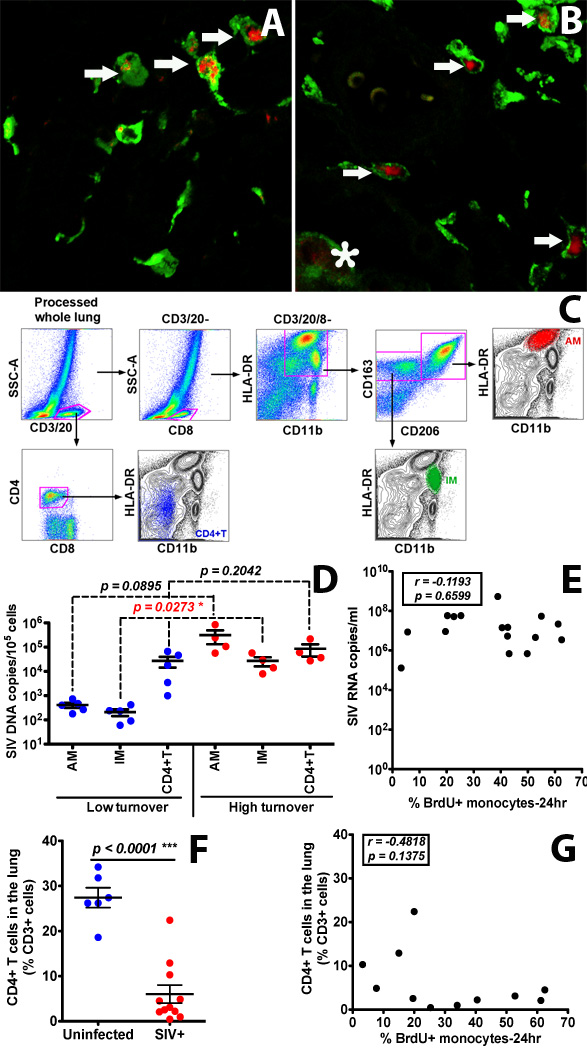
Figure 6. Infection and replication of SIV in IM and AM contribute to the viral load in lung tissues of infected rhesus macaques exhibiting high monocyte turnover.

(A&B) Confocal microscopy was performed on lung tissues obtained from SIV-infected monkeys with low (≤ 30%) monocyte turnover (A: n=3) and higher (> 30%) blood monocyte turnover levels (B: n=6). Anti-CD163 (Green) antibody was used to identify macrophages, and SIV RNA (Red) was detected with anti-sense riboprobes. Arrows indicate SIV-infected IM. The asterisk indicates an SIV-infected AM. (C) Gating strategy for analyzing and sorting AM, IM and CD4+ T cells isolated from lung tissue. (D) IM, AM and CD4+ T cells were sorted via FACS from single-cell suspensions of lung tissues from SIV-infected monkeys with low (n=5) and high (n=4) blood monocyte turnover, and SIV DNA levels were quantitated and standardized against RNase P levels. (E) Plasma VL did not correlate with monocyte turnover in SIV-infected macaques. (F) CD4+ T cell depletion in the lung of all SIV-infected macaques was evident (p<0.0001). (G) The levels of CD4+ T cells did not correlate with monocyte turnover rates in SIV-infected macaques. Student’s t test was applied for comparisons in results shown in panels D and F, and Spearman’s correlation analysis was applied in results shown in panels E and G.
