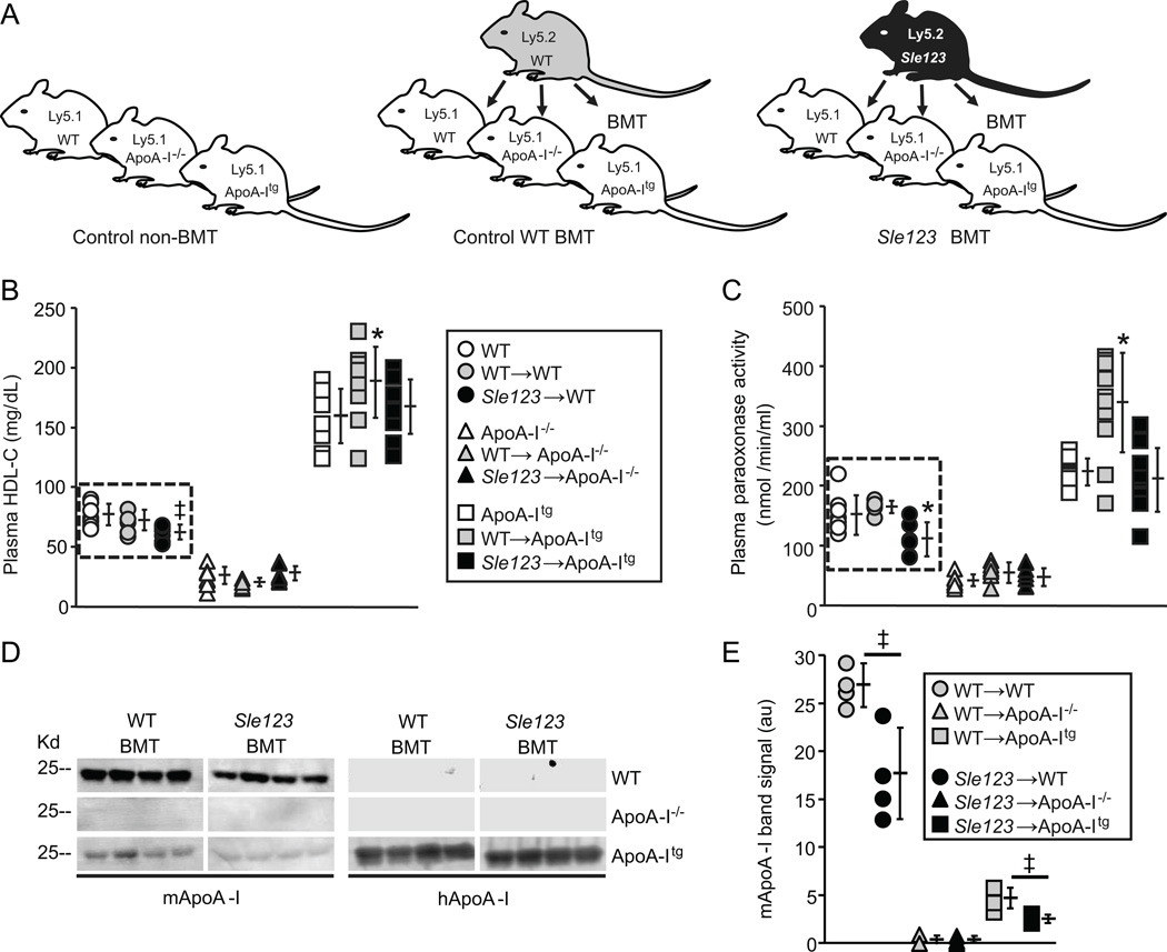
Figure 1.
(A) Schematic of BMT scheme used to generate chimeric autoimmune mice (animals were transplanted with WT or Sle123 BM at 6 weeks of age). (B) Plasma HDL-C levels in the indicated control, control WT BMT and Sle123 BMT mice (38 weeks of age, 32 weeks post-BMT, n=8). (C) Plasma PON1 activity against paraoxon substrate in the indicated control, control WT BMT and Sle123 BMT mice (38 weeks of age, 32 weeks post-BMT, n=8). Note autoimmune-mediated reductions in HDL-cholesterol (HDL-C) and HDL-associated PON1 activity in Sle123→WT mice (in dashed box). (D) Plasma levels of mouse ApoA-I (mApoA-I) and human ApoA-I (hApoA-I) in 4 control and 4 Sle123 BMT mice (38 weeks of age, 32 weeks post-BMT). Graph showing band intensities for plasma mApoA-I is shown in (E). Note reduction in plasma mApoA-I in Sle123→WT mice compared to their control WT counterparts. ApoA-Itg animals have markedly lower endogenous mApoA-I levels as previously reported (19). (All data mean ± SD, *p<0.05 by one way ANOVA within WT, ApoA-I−/− or ApoA-Itg group, ‡p<0.05 by T test compared to control group).

