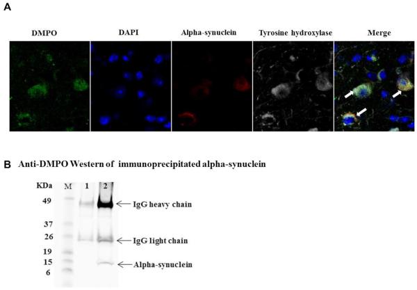
Figure 3.
Detection of alpha-synuclein-centered radicals in dopaminergic neurons. (A) Confocal images of cryocut midbrain sections from sham + DMPO- and Maneb + Paraquat (MP) + DMPO-treated mice showing the colocalization of anti-DMPO staining with alpha-synuclein and tyrosine hydroxylase. (B) Immunoprecipitation of DMPO adducts. Proteins were immunoprecipitated from nigrostriatal tissue homogenates using either normal mouse IgG as a control (lane1) or with anti-alpha-synuclein antibody followed by Western blot analysis of immunoprecipitates using rabbit anti-DMPO antibody. Blots are representative of four different immunoblots from an equal number of experiments.

