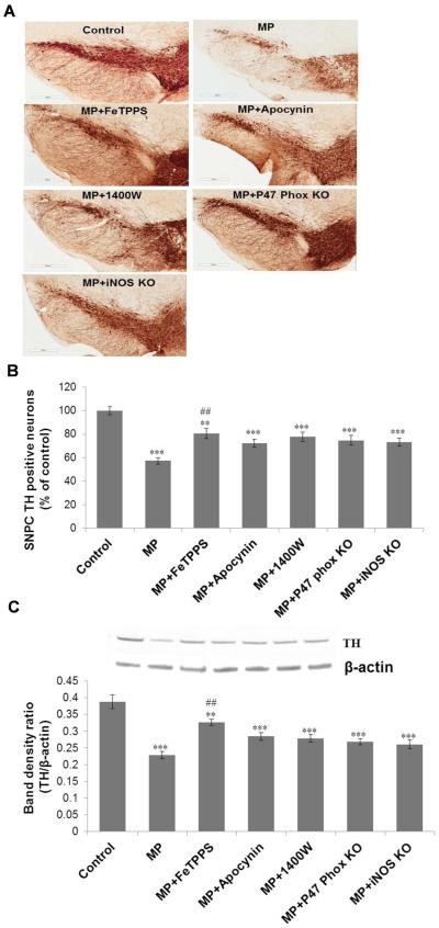
Figure 6.
Tyrosine hydroxylase (TH) immunoreactivity in the substantia nigra of mouse brains following 6 weeks of Maneb and paraquat (MP) coexposure. (A) Upper panel shows TH immunoreactivity in frozen brain sections of control and treated animals with or without pretreatment with FeTPPS (10mg/kg, i.p.), apocynin (10mg/kg, i.p.), or 1400W (15 mg/kg, i.p.), and in p47 phox or iNOS knockout mice. (B) Lower panel shows the number of TH positive neurons in the substantia nigra pars compacta (SNPC) region of all experimental groups as in Figure A. (C) Western blot analysis of TH protein expression in the nigrostriatal tissue of mouse brains following 6 weeks of Maneb and paraquat coexposure in the presence and absence of pretreatments as indicated in the graph. The upper panel depicts a representative blot of TH protein; the lower panel depicts a densitometric analysis of the same with β-actin as the reference. The data are expressed as mean ± SEM (n=3–5). (** p<0.01, ***p<0.001 as compared with control and ## P<0.01, as compared with the Maneb and paraquat coexposed group.)

