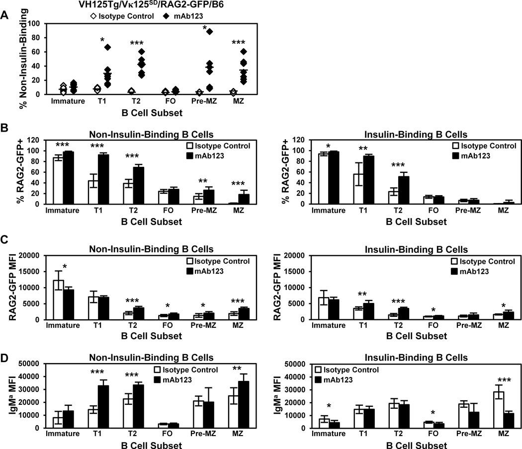
Figure 7. Targeting insulin-occupied BCR increases RAG2-GFP expression and the frequency of non-insulin-binding B cells in developing subsets.
VH125Tg/Vκ125SD/RAG2-GFP/B6 7–17 week old mice were injected i.p. with 100 µg anti-insulin mAb123 (black, n = 7) or isotype control mAb (white, n = 6) i.p. every 2 days for 1 week. Bone marrow and spleens were freshly isolated 1 d after the final injection and B cell subsets were identified as in Fig. 1D. (A) The frequency of non-insulin-binding B cells in individual mice is plotted, bars indicate the average. (B–D) The average percentage of RAG2-GFP+ cells (B), the average RAG2-GFP MFI of RAG2-GFP+ cells (C), or the IgM MFI (D) of non-insulin-binding B cells (Left) or insulin-binding (Right) in each subset is shown, error bars indicate SD. * p < 0.05, ** p < 0.01, *** p < 0.001, two tailed t test.

