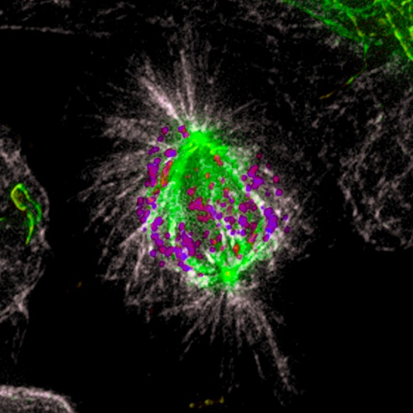
Figure 1. Microtubule detyrosination guides chromosomes to the spindle equator.

Immunofluorescence of a methanol fixed U2OS cell in mitosis. Total α-tubulin in white; detyrosinated tubulin in green; kinetochores (ACA) in purple. Note that the astral microtubules are not detyrosinated.
