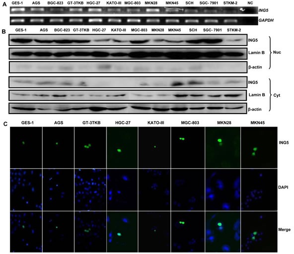
Figure 1. ING5 expression and localization in gastric cancer and epithelial cell lines.

A: Different amplicons of ING5 mRNA were detected and showed inconsistent density in all gastric cancer and epithelial cells with an internal control of GAPDH. B: Cytosolic (Cyt) and nuclear (Nuc) fraction proteins were loaded and probed with anti-human ING5 antibody (28kDa) with β-actin (42 kDa) or Lamin B (60kDa) as an internal control. C: GES-1, AGS, GT-3TKB, HGC-27, KATO-III, MGC-803, MKN28 and MKN45 cells were transiently transfected with pEGFP-N1-ING5 plasmid and observed under confocal microscope (green for ING5; DAPI, blue for nucleus). NC, negative control.
