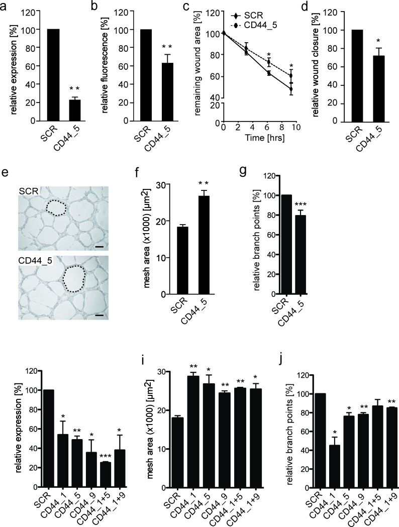
Figure 4. Reduced CD44 membrane expression on EW7 cells decreases tumor cell migration and vasculogenic network formation.
A. qRT-PCR and B. flow cytometic analysis of CD44 mRNA and protein expression, respectively, 48 hours after transfection of VM+ EW7 Ewing sarcoma cells with non-silencing (SCR) or CD44 targeting (CD44) siRNA. C. Migration analysis of transfected EW7 cells on hyaluronic acid. Relative wound closure is displayed as the percentage compared to T = 0. D. Relative wound closure by transfected EW7 cells as compared to SCR after 6 hours. E. Representative images of vasculogenic networks formed on Matrigel by SCR and CD44 targeted transfected cells. Network meshes are indicated by a dotted line. Scale bars represent 200 μm. F. Quantification of the average network mesh area in networks formed by SCR or CD44 transfected EW7 cells (n = 3). G. Quantification of the relative percentage of branch points. H. qRT-PCR analysis, 48 hours after transfection of VM+ EW7 Ewing sarcoma cells with non-silencing (SCR) or different CD44 targeting siRNAs (CD44_1, CD44_5, CD44_9) and combinations thereof (CD44_1+5, CD44_1+9). I. Quantification of average network mesh area and relative percentage of branch points J. of EW7 cells transfected with SCR or different CD44 siRNAs. Data are presented as mean ± SEM. *p < 0.05; **p < 0.01; ***p > 0.001.

