
Figure 1. COP1 directly interacts with p27.
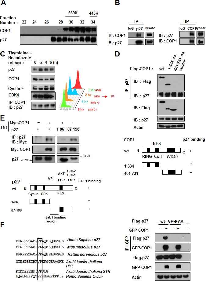
(A) COP1 and p27 coeluted as part of the high molecular weight complex. Gel filtration and elution profiles analysis of COP1 and p27. The distributions of these proteins were analyzed by gel filtration chromatography (Superose 6). Immunoblots of the fractions for indicated proteins are shown in U2OS cells. Molecular size of eluted fraction is indicated above. (B) Endogenous interaction of COP1 and p27 was observed. Lysates of U2OS cells were prepared and equal amounts of cell lysates were analyzed by immunoprecipitation (IP) with either control mouse IgG or p27 and analyzed by immunoblotting (IB) with anti-COP1. Lysates were also analyzed by IP with the indicated antibodies and IB with anti-p27. (C) p27 and COP1 interacted during the cell cycle. U2OS cells were synchronized to the G2/M phase using treatment with thymidine-nocodazole. Lysates of synchronized cells were analyzed by IB with the indicated antibodies. COP1-p27 interaction at various phases of the cell cycle was detected by IP with the COP1 antibody followed by IB with anti-p27 (left). Cell samples at labeled time points after release of nocodazole were stained with propidium iodide and analyzed by FACS for DNA content. DNA content histograms are shown for the time points indicated (right). (D) p27 bound to the N-terminus of COP1 but not to the C-terminus. Wild-type (wt; aa 1-731), N-terminal (aa 1-334), or C-terminal (aa 401-731) Flag-COP1 was transfected into HeLa cells. Cells were treated with MG132, and cell lysates were analyzed by IP with anti-p27 and IB with anti-Flag. (E) COP1 bound to the C-terminus of p27. Myc-COP1 and PET-p27 were transcribed and translated in vitro (TNT). COP1 and p27 proteins were incubated overnight and analyzed by IP with anti-p27 followed by IB with anti-Myc. (F) The interaction of COP1 and p27 was mediated by the conserved VP sequence on p27. p27 has the VP motif for COP1 binding. Consensus COP1 binding motif, highlighted in sequences of p27 in human and other species' DNA for comparison. HY5, STH, and c-Jun proteins are known COP1 binding proteins with VP motif. 293T cells were transfected with the indicated plasmids and treated with MG132. Cell lysates were analyzed by IP with anti-GFP and IB with anti-Flag.