Fig. 1.
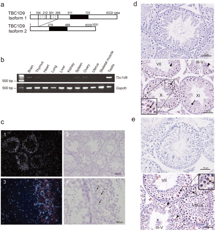
Analyses of TBC1D9 expression in 8-week-old mouse tissues. (a) Scheme of mouse TBC1D9 isoforms. Isoform 1 protein (1264 amino acids) possesses two GRAM domains (dotted box). A TBC domain (solid box) exists in both isoform 1 and isoform 2 (1031 amino acids). The open bars represent regions amplified by RT-PCR. (b) RT-PCR analysis of Tbc1d9 and Gapdh mRNA in mouse tissues. Top, amplification of both isoforms of Tbc1d9; bottom, amplification of Gapdh. (c) In situ hybridization of Tbc1d9 mRNA in the mouse testis. Light microscopic dark (1, 3) and light field (2, 4) images of Tbc1d9 mRNA in sections of the testis. Signal is seen in the cytoplasm of spermatocytes (arrows). (d) and (e) Immunohistochemical analysis of TBC1D9 proteins in the mouse testis using anti-mouse TBC1D9 antiserum (d) and anti-human TBC1D9 antibody (e). Top, negative control; bottom, immunoreactivity is seen in the cytoplasm of spermatocytes (arrows) and round spermatids (arrowheads). Roman numerals, the stages in the seminiferous tubules.

