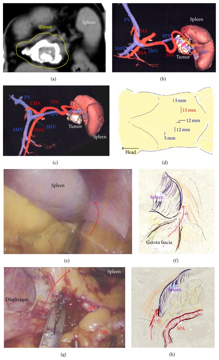
Figure 1.
(a) The tumor surface was severely calcified (yellow circle). (b) Three-dimensional images revealed that the tumor involved the SPV (yellow arrows). (c) The SPA was not involved in the tumor. (d) A total of five ports were placed. (e) and (f) The inferior ligament of the spleen was cut (red arrow). (g) and (h) The superior ligament of the spleen was cut (red arrow). SPA, splenic artery; SPV, splenic vein.

