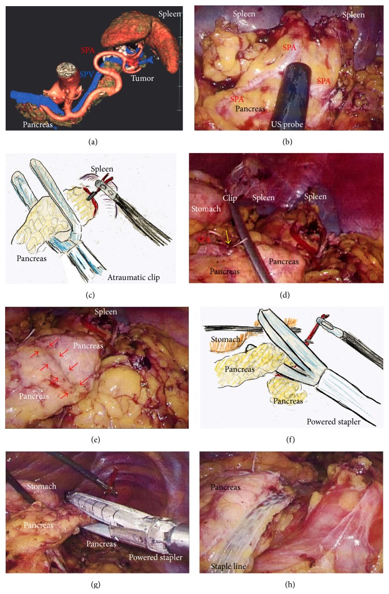
Figure 3.
(a) Tumor location and anatomical landmarks were assessed preoperatively. (b) Ultrasound was performed to confirm the tumor location and to determine the cutting line. (c) Compression using an atraumatic clip was required before proceeding further because stapling was performed using a Covidien stapler. (d) The proximal SPA was ligated (yellow arrow) and the pancreas was compressed. (e) Compressed parenchyma was confirmed before proceeding further (red arrows). (f), (g), and (h) The pancreas and SPV were cut en bloc using the powered stapler; the pancreas was retraced with tape. SPA, splenic artery; SPV, splenic vein.

