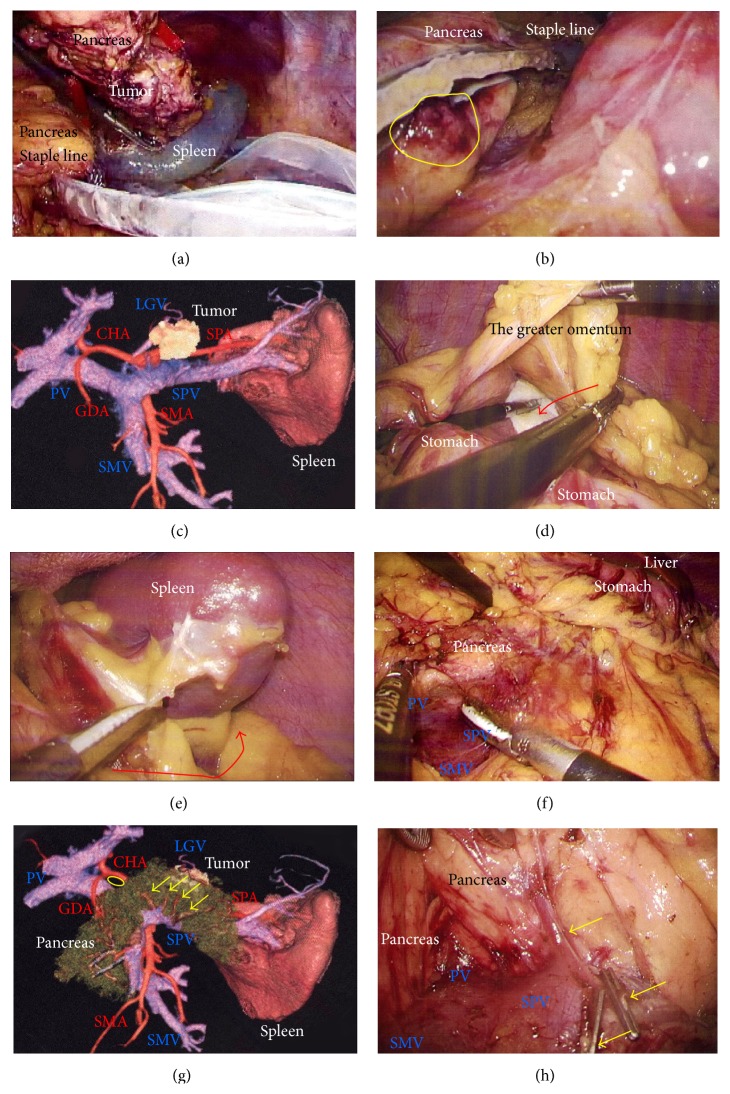
Figure 4.
(a) The specimen was extracted through a 3 cm incision in an endobag to prevent dissemination by direct contact with the tumor. (b) The pancreatic stump and capsule were checked carefully. Subtle injury to the pancreatic membrane caused postoperative leakage of pancreatic fluid (yellow circle). (c) The tumor location and invasive signs were assessed carefully. (d) Gauze was placed on the splenic hilus as a landmark. The greater omentum was then cut (red arrow), and the omental bursa was opened. The left gastroepiploic vessels were cut. (e) The splenocolic ligament along the inferior spleen was then cut (red arrow). It was critical that the reverse side of the thin membrane of the transverse mesocolon was confirmed beforehand, via a bird's-eye view. (f) The confluence of the SMV, SPV, and PV was confirmed. (g) Branches from the SPV were detected before proceeding further (yellow arrows). A suitable dissection point for CHA skeletonization was also determined preoperatively (yellow circle). (h) Venous branches from the SPV were then skeletonized (yellow arrows). These venous branches were singly clipped and then cut by LCS. CHA, common hepatic artery; LCS: laparoscopic coagulation sheers; PV, portal vein; SMV, superior mesenteric vein; SPV, splenic vein.

