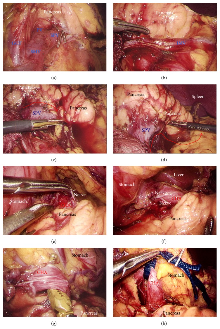
Figure 5.
(a) The GCT was visualized. (b) The IMV was then skeletonized and preserved. (c) The SPV was skeletonized from the pancreatic parenchyma (red arrow). (d) The SPV was separated from dorsal fixation by connective tissue (red arrow). (e) and (f) The nerve surrounding the arterial sheath was useful to grasp the CHA without arterial injury. (g) and (h) The CHA was skeletonized and taped. CHA, common hepatic artery; GCT, gastrocolic trunk; IMV, inferior mesenteric vein; SPV, splenic vein.

