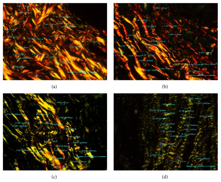
Figure 3.
Photomicrograph of PSR stained sections under polarized light microscopy (40x). (a) NM showing predominantly thick YO collagen fibres. (b) WDSCC showing predominantly thick YO and thin GY collagen fibres. (c) MDSCC showing thick YO and thick GY collagen fibres. (d) PDSCC showing thick GY and thin GY collagen fibres.

