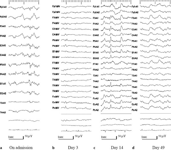
Fig. 2.

EEGs. a On admission: bilateral periodic sharp waves of approximately 0.5 Hz with the right amplitude slightly higher than the left. b Day 3: sharp waves with maximum amplitude at the right temporal area. c Day 14: occasional spikes in the left posterior quadrant with right hemispheric suppression. d Day 49: no apparent spikes. The recording of day 3 was averaged reference montage. Otherwise the recordings were ear reference montage.
