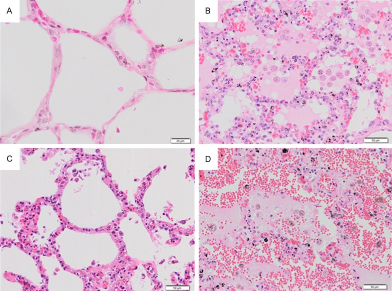
Figure 1.

Histopathological changes in H&E-stained lung tissues of severe falciparum malaria patients. Normal lung tissue (A). The lung tissue of severe falciparum malaria patient showing edema fluid in the alveoli (B). The alveoli are filled with PRBCs, RBCs, and pigment-laden macrophages. PRBCs sequester in the septal capillaries and small blood vessels in the lung of severe falciparum malaria (C). Alveolar hemorrhage was always seen in the lung tissues of severe falciparum malaria patients (D). All images are 200 × magnification. Bar = 50 μm.
