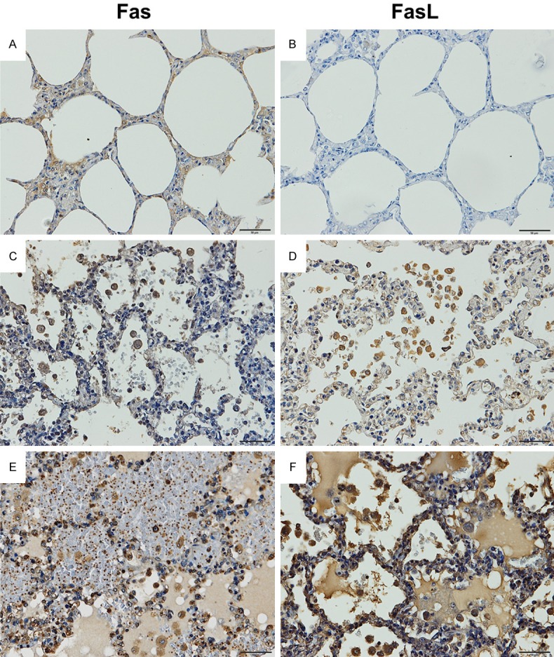
Figure 2.

Representative results of immunoperoxidase staining for Fas and FasL in lung tissue of severe falciparum malaria patients. Normal lung tissue (A and B). The lung tissue of a severe falciparum malaria patient with non-PE (C and D). The lung tissue of a severe falciparum malaria patient with PE (E and F). All images are 200 × magnification. Bar = 50 μm.
