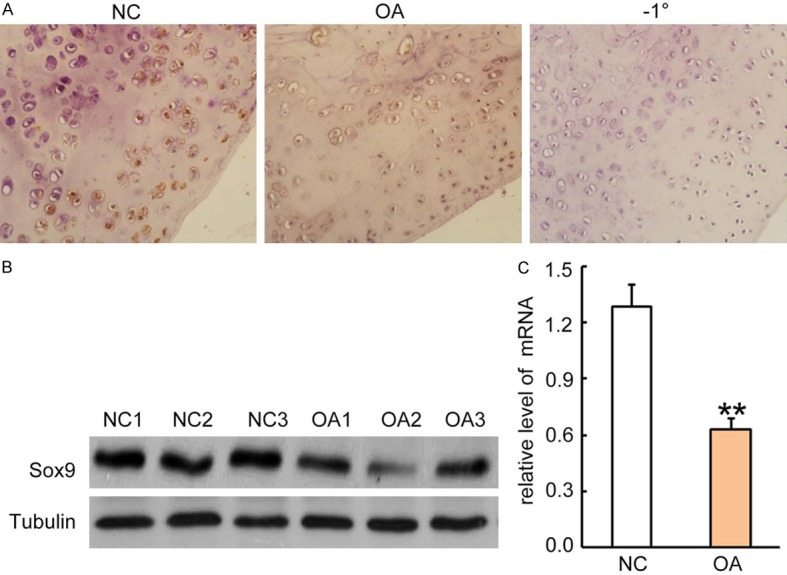
Figure 1.

Sox9 expression was reduced in OA patients. A. Immunohistochemical staining for Sox9 expression in articular cartilage tissues from patients with OA and normal controls. Rabbit anti-human Sox-9 antibody (1:500 dilution) was incubated with sections for 2 hrs and followed by incubation with HRP-conjugated second antibody for 1 hour. The Sox9 positive staining was developed by the addition of DAB substrate. Sections from normal controls were incubated with second antibody only as a negative control (right panel). One representative photograph of two groups is shown. B. Western blotting analysis for Sox9 expression in cell lysates from articular cartilage tissues. Each lane represents a sample from individual human subjects. After stripping, the GAPDH antibody was incubated for sample loading controls. C. Sox9 mRNA levels in articular cartilage tissues were quantitatively analyzed by qRT-PCR. Data was normalized by internal control GAPDH and presented as mean DDCt relative to GAPDH ± standard error, n = 3. *P < 0.05, **P < 0.01.
