
Figure 6.
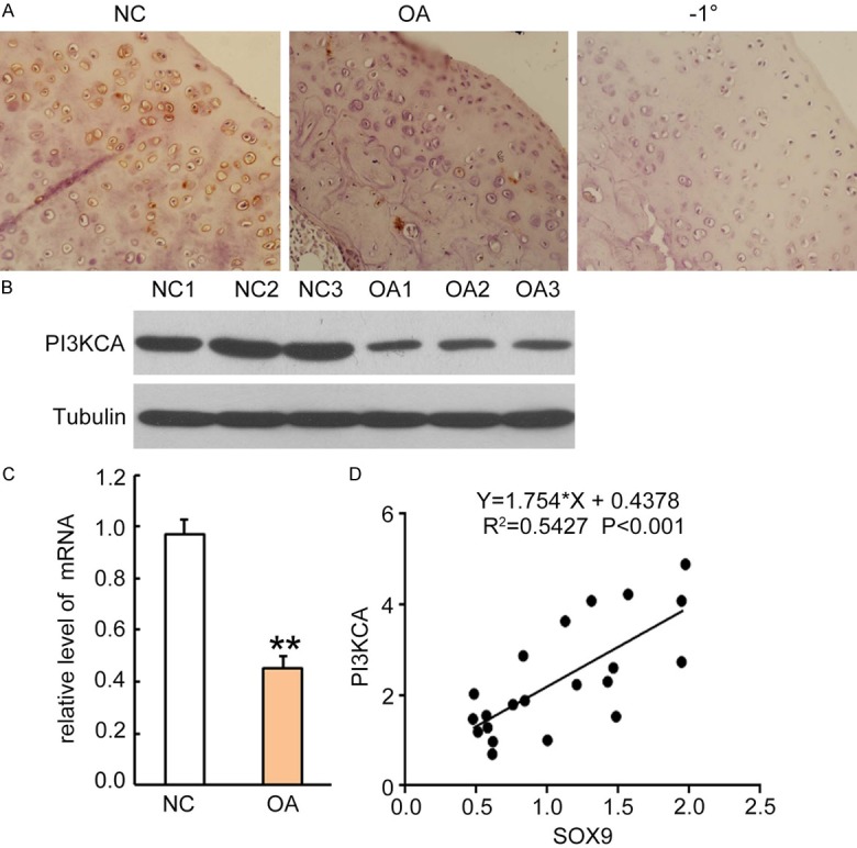
Sox9 expression level was positively correlated to PI3KCA in articular cartilage tissues from patients with OA. A. Rabbit anti-human PI3KCA antibody (1:300 dilution) was incubated on sections for 2 hours and followed by incubation with HRP-conjugated second antibody for 1 hour. The PI3KCA positive staining was developed by addition of DAB substrate. Sections from normal controls were incubated with second antibody only as negative control (right panel). One representative photograph of each group sample is shown. B. Western blotting analysis for PI3KCA expression in cell lysates from articular cartilage tissues of normal subjects and patients with OA. Each lane represents sample from an individual human subject. After stripping, internal control antibodies against tubulin were incubated for detection of internal control proteins. C. PI3KCA mRNA was quantitatively analyzed by qRT-PCR. Data was normalized by internal control GAPDH and presented as mean DDCt relative to GAPDH ± standard error, n = 3. *P < 0.05, **P < 0.01. D. Correlations between Sox9 and PI3KCA was plotted as a scatter plot, and statistical significances were analyzed by the R-squared test, *P < 0.05 is considered as significant regression.