Figure 1.

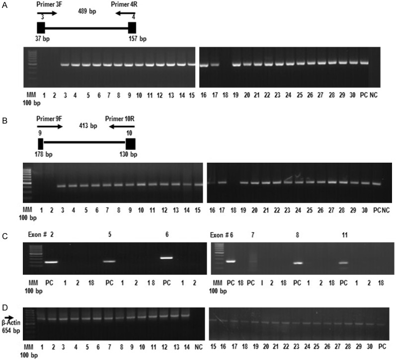
The FBXW12 gene is deleted in some EOCs. (A) Gel image showing the absence of a 489 bp PCR product in three tumor samples (1, 2 and 18), after amplification of ovarian genomic DNA, using primers targeting exons 3 and 4 and the corresponding intronic sequence of the FBXW12 gene. (B) Gel image showing the absence of PCR products following amplification of ovarian genomic DNA from tumors 1, 2 and 18, using primers targeting a 413 bp fragment comprising exons 9 and 10 and the corresponding intronic sequence of FBXW12. (C) Gel image showing the absence of PCR products derived from tumors 1, 2 and 18, using primers targeting the remaining 6 exons (2, 5, 6, 7, 8 and 11) of the FBXW12 coding sequence. (D) The lack of PCR products in tumors 1, 2 and 18 is in contrast to the amplification of a 654 bp fragment of a constitutive gene (β-actin) from the same tumor samples. Genomic DNA isolated from white cells of healthy women was used as positive control (PC). Also in all PCR experiments, a reaction with all PCR components with the exception of DNA was used as negative control (NC). MM = molecular marker, PC = positive control, NC = negative control, 1 = stage III serous EOC with complete deletion of FBXW12 coding region, 2 = stage IV serous EOC with complete deletion of FBXW12 coding region, 18 = stage III serous EOC with complete deletion of FBXW12 coding region, 3-30 = EOCs of different histotypes with no deletion of FBXW12 coding region. The diagrams on top of panels (A) and (B) depict the approximate positions of the primers used for amplification.
