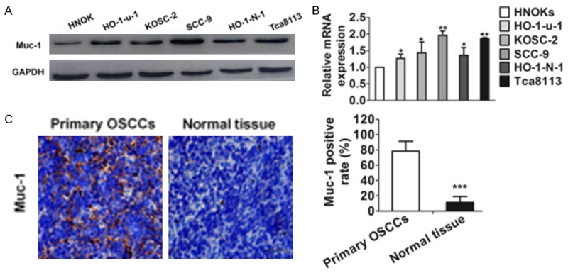
Figure 1.

Evaluation of muc-1 expression in OSCC-derived cell lines. A. Immunoblotting analysis of muc-1 protein in the OSCC-derived cell lines and HNOK. Muc-1 protein expressions are up-regulated in all OSCC-derived cell lines examined compared with that in the HNOK. B. Quantification of muc-1 mRNA levels in OSCC-derived cell lines by qRT-PCR analysis. All OSCC-derived cell lines have significant up-regulation of muc-1 mRNA compared with that in the HNOK (*P<0.05 and **P<0.01 versus HNOK). C. Evaluation of muc-1 protein expression in primary OSCCs. Representative IHC results for muc-1 protein in normal tissue and primary OSCCs. Strong muc-1 immunoreactivity is detected in primary OSCCs. Normal oral tissues show almost weak immunostaining. Scale bars, 50 μM. Bars are represented as the mean ± SD, n=3, *P<0.001 versus normal tissue).
