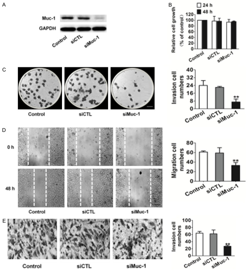
Figure 2.

Effects of muc-1 silencing on SCC-9 cell clone formation motility. A. Immunoblotting analysis shows that the muc-1 protein levels in siMuc-1 cells have decreased markedly compared with that in mock cells. B. Proliferation assays indicated that muc-1-siRNA exerted no inhibition on SCC-9 cells proliferation in vitro. C. Clone formation assay indicated that muc-1 siRNA inhibited SCC-9 cell colony-forming effectively. Scale bars: 1 mm. D. Representative examples of wound healing experiment results from control and siRNA-muc-1 SCC-9 cells. Representative pictures of the wound distance were taken at each time point as indicated. Scale bars: 50 µm. E. SCC-9 motility was also estimated by Transwell assay. Representative pictures were taken after staining with crystal violet. Scale bars: 50 µm. Invaded cells transfected with scrambled siRNA or muc-1-siRNA were evaluated on the basis of the mean values from five fields for each group (Bars are represented as the mean ± SD, n=3, **P<0.01 versus control cells).
