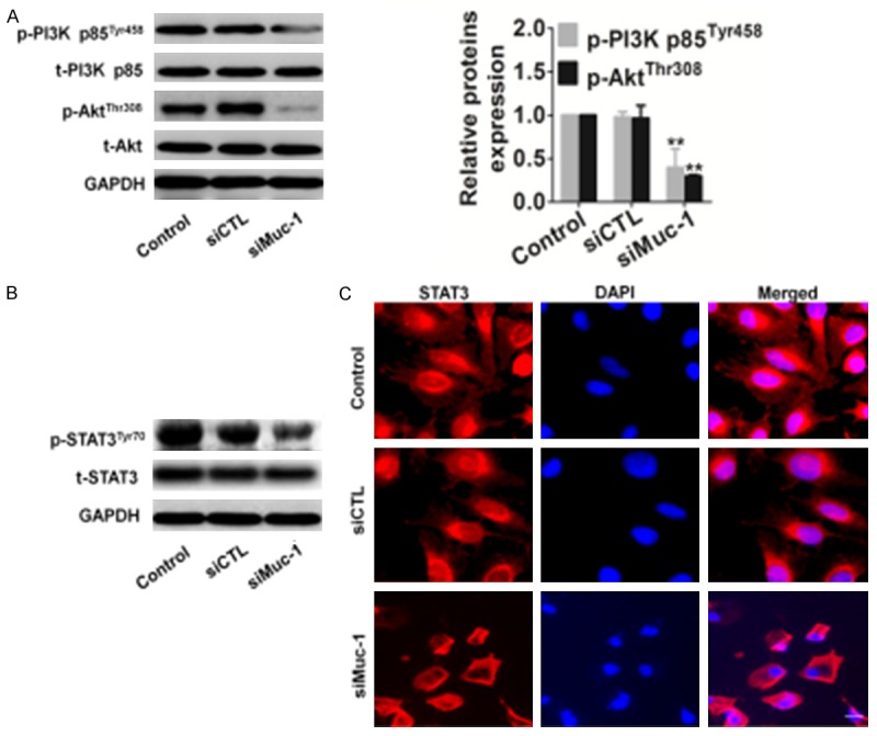
Figure 3.

Effects of muc-1 silencing on the PI3K/Akt pathway. A. Expression of PI3K and Akt in muc-1-silenced cells. After transfection of cells with either scrambled siRNA or muc-1-siRNA, the total cells were isolated and PI3K/Akt expression levels were detected by western blotting. GAPDH was used as a loading control. B. SCC-9 cells transfected using the same conditions as described above and total protein was extracted, the total and phosphorylated (p) STAT3 were detected. C. SCC-9 cells were transfected, and then cells were fixed and incubated with primary antibody against STAT3. SCC-9 cells were immunostained with anti-rabbit FITC-conjugated secondary antibody and then stained with DAPI. The specimens were visualized and photographed using a fluorescence microscope. Scale bars: 20 µm. Blue depicts the nucleus and green depicts localization of STAT3 (Bars are represented as the mean ± SD, n=3, **P<0.01 versus control cells).
