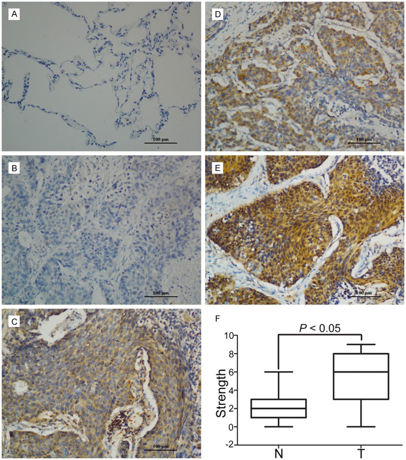
Figure 2.

Expression of E2F2 in N SCLC tissues by IHC. A. Micrographs showed the staining of E2F2 in normal lung tissues. B-E. Micrographs showed the staining of E2F2 in tumor tissues (negative, weak, modern, strong). F. Reproducibility of the measurement in all 86 patients was calculated using the Wilcoxon matched paired test.
