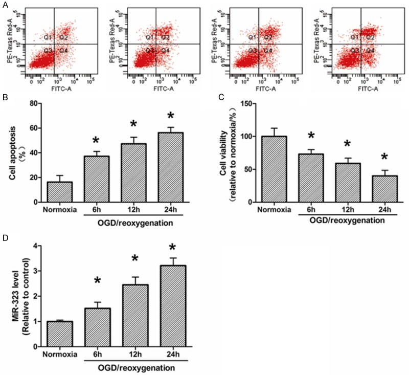
Figure 1.

Apoptosis, survival and expression of miR-323 in hippocampal neurons after OGD-induced ischemic injury. A. Flow cytometry analysis of neuronal apoptosis during 6-24 h of reoxygenation post-3 h OGD. B. Quantization of A. C. XTT analysis of neuronal survival during 6-24 h of reoxygenation post-3 h OGD. D. The expression of miR-323 in neurons during 6-24 h of reoxygenation post-3 h OGD. The data presented as mean ± SD. *P<0.05 compared to the sham group. N=3, *P<0.05, compared with Normoxia.
