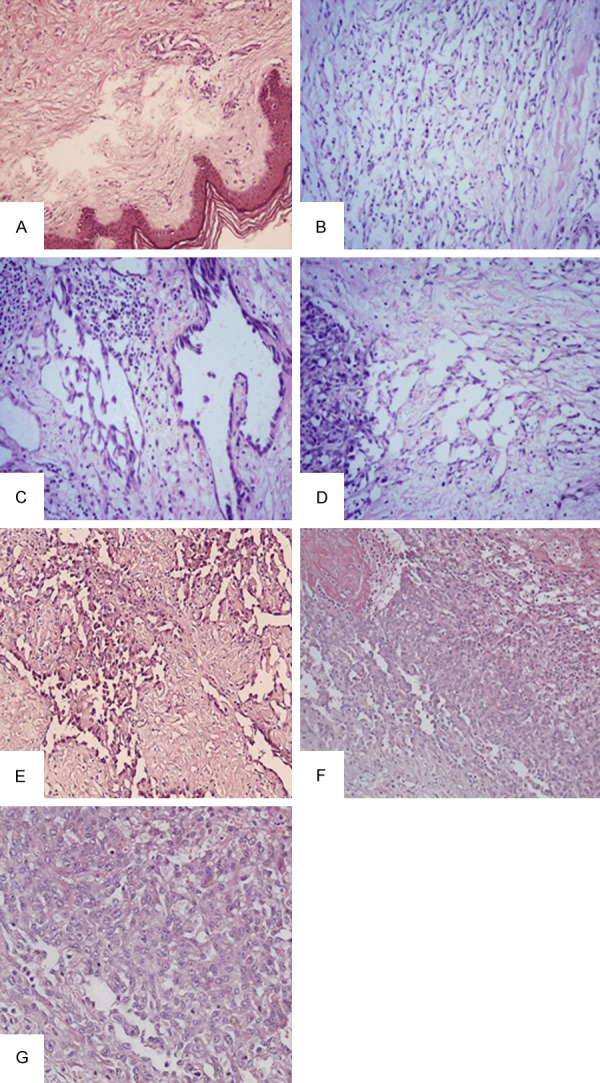
Figure 2.

A: Hyperkeratosis of the epidermis, mild lymphocytic infiltrate in the upper part of the dermis, and varying degrees of edema in the interstitial tissues. (HE × 200); B: Marked lymphangiomatosis (HE × 200); C: Endothelial hyperplasia in the subcutaneous tissues (HE × 200); D: Marked lymphangiomatosis adjacent to the area of frankly malignant disease. (HE × 200); E: Well differentiated areas showed luminal pattern with numerous islands and cords of connective tissue surrounded by one to several layers of malignant cells. (HE × 200); F: Undifferentiated areas, the cells were arranged in a solid pattern. They were large, of epithelioid type, The cytoplasm was sparse, slightly eosinophilic. Between the cells was erythrocytes “lake”. (HE × 200); G: Many mitotic figures were seen. (HE × 400).
