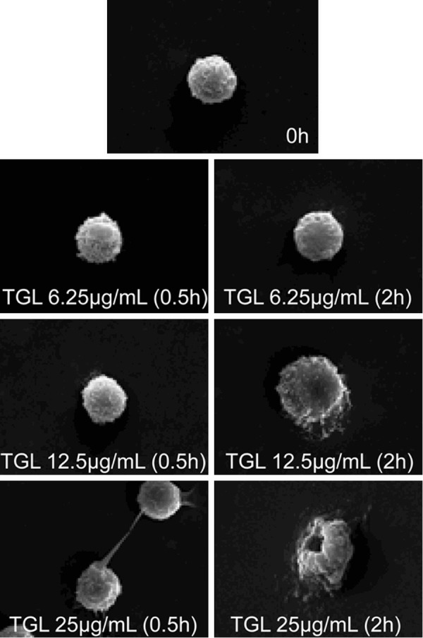
Figure 1.

Surface morphology change of macrophage. SEM images showing that After using 6.25 μg/mL purified TGL to stimulate macrophage for 0.5 h and 2 h respectively, there was no evident change in the morphology of macrophage, but after using 12.5 μg/mL to stimulate macrophage for 2 h, the macrophage increased in volume, evident wrinkles were found on surface, after using 25 μg/mL to stimulate macrophage for 0.5 h, pseudopod was produced on cell surface, and it died 2 h later owing to cytomembrane damages.
