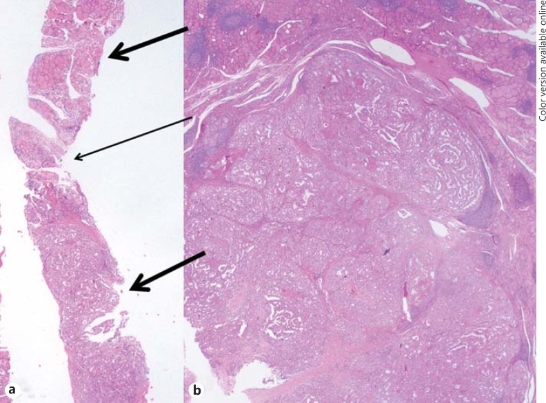
Fig. 2.
CNB histology and surgical pathology of a papillary thyroid carcinoma. a This CNB histology specimen shows tumor cells with an atypical papillary growth pattern (lower thick arrow), diagnosed as papillary thyroid carcinoma. The specimen also shows a normal gland (upper thick arrow) and intervening fibrous capsule-like structure between the tumor and normal gland (thin arrow). b Histology of the surgical specimen demonstrates tumor cells and an adjacent normal gland corresponding to the histology of CNB, diagnosed as papillary thyroid carcinoma. The diagnosis of the initial and repeat FNA was NA (not shown). HE stain. ×10.

