Fig. 4.
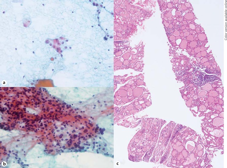
FNA cytology and CNB histology of focal lymphocytic thyroiditis manifested as a thyroid nodule. a The initial FNA cytology shows a few scattered atypical follicular epithelial cells with nuclear enlargement, size variation and pale chromatin, with the diagnosis of NA. b The repeat FNA cytology shows some atypical follicular epithelial cells with enlarged overlapping nuclei, occasional nuclear grooves, pale chromatin and some lymphocytes, repeatedly diagnosed as NA. c The CNB histology specimen shows variable-sized follicles with mild oncocytic change, and lymphocytic infiltration (arrow) without capsulation, diagnosed as lymphocytic thyroiditis. HE stain. ×10.

