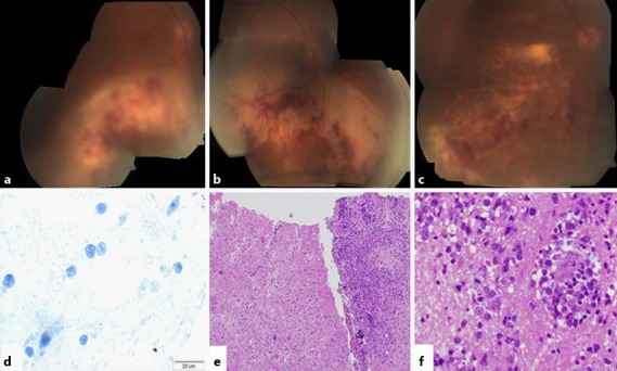
Fig. 1.

Posterior segment presentation and histopathological examination. a Vitritis and a white-yellow lesion with few retinal and subretinal hemorrhages in the inferior retina, without macular or optic disc involvement. b The inferior lesion advanced to the inferior border of the optic disc and involved the macula but not the fovea. c The subretinal lesion crossed the fovea and advanced to the temporal border of the optic disc. d CD3 staining showed strongly immunoreactive cells. e Brain biopsy. f CNS lymphoma: large lymphocytes with giant nucleus, open chromatin and few mitoses.
