Fig. 3.

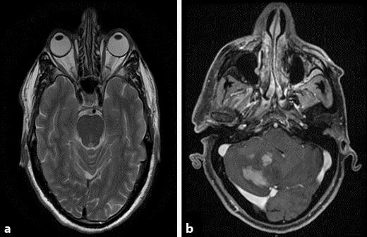
MRI. a T2 axial cross-section was normal. b T2 axial cross-section showing several new space-occupying lesions in the right cerebellum.

MRI. a T2 axial cross-section was normal. b T2 axial cross-section showing several new space-occupying lesions in the right cerebellum.