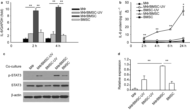
Figure 1.
Juxtacrine interaction of BMSCs and macrophages (MФ) induced IL-6 activation. (a) Real-time PCR revealed significantly higher levels of IL-6 mRNA at 2 and 4 hours in co-cultures of mouse primary macrophages and BMSCs compared to co-cultures of macrophages and apoptotic BMSCs (UV treated), or macrophage and BMSC cultures alone. Data are mean ± SEM (n = 2 in each group); **P < 0.01. (b) ELISA assay showed significantly higher levels of IL-6 protein in the supernatant from co-culture of mouse primary macrophages and BMSCs at 2, 4, 6, and 24 hours compared to co-culture of macrophages and apoptotic BMSCs, or macrophage and BMSC cultures alone. Data are mean ± SE (n = 2 in each group); *P < 0.05; **P < 0.01. (c) Western blot analysis showed endogenous STAT3 phosphorylation was increased in co-cultures of primary macrophage and BMSCs after 6 hours when compared with either co-culture of macrophages and apoptotic BMSCs, or BMSCs, apoptotic BMSCs and macrophages alone. (d) Quantification for phosphorylated STAT3 protein expression relative to total STAT3. Data are mean ± SEM of two independent experiments (n = 2 in each group); **P < 0.01.

