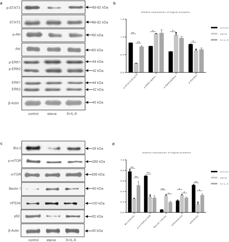
Figure 3. Inhibition of starvation-induced autophagy by IL-6 was STAT3- dependent.
(a) STAT3, Akt and ERK1/2 were analyzed by immunoblotting under starvation conditions and incubation with or without IL-6 (30 ng/mL) for 2 h. β-actin was used as the loading control. (b) Relative expression levels of STAT3, Akt and ERK1/2 were shown in accordance with immunoblot analysis in Fig. 3a. Control versus starve, starve versus S+IL-6; *P < 0.05, **P < 0.01. (c) Immunoblot analysis of mTOR, Bcl-2, Beclin 1, VPS34 and p62 in cells treated with or without IL-6. (d) Relative expression levels of mTOR, Bcl-2, Beclin 1, VPS34 and p62 were shown in accordance with immunoblot analysis in Fig. 3c. Control versus starve, starve versus S+IL-6; *P < 0.05, **P < 0.01.

