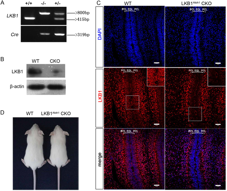
Figure 1. Generation of LKB1Atoh1 CKO mice.
(A) Mouse genotyping by PCR analysis. Lanes: wild-type (+/+), homozygous (−/−) and heterozygous (+/−) mice. (B) Western blot of Lkb1 in the cerebellum of P14 LKB1Atoh1 CKO and wild-type mice. A 48.6-kD LKB1 protein was obviously decreased in the cerebellum of LKB1Atoh1 CKO mice (CKO) compared to the controls (WT). (C) Expression pattern of LKB1 in the P3.5 cerebellum. Confocal images of the cerebellum in wild-type and LKB1Atoh1 CKO mice. In the P3.5 wild-type cerebellum, LKB1 was primarily localized throughout the entire lobe, including the PCL and EGL (parentheses) which mainly consist of the granule cell precursors (GCPs), while in the LKB1Atoh1 CKO cerebellum, LKB1 expression was not detected in the GCPs of the ECL. Inserts represent high magnification view of the boxed areas. Highly magnified images confirmed no detectable expression in the GCPs of the EGL in mutant mice. Red, LKB1; Blue, DAPI. Scale bar: 50 μm. (D) Gross morphology of P21 wild-type and LKB1Atoh1 CKO mice. There were no obvious differences, except for the smaller body size of the mutant mice compared to the controls.

