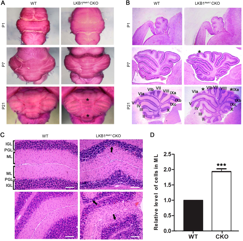
Figure 3. Morphological abnormalities of the cerebellum in the mutant mice.
(A) Abnormal volume of the cerebella in the LKB1Atoh1 CKO mice. At P1 and P7, the volume of the LKB1Atoh1 CKO cerebellum was similar to the wild-type. However, the P21 mutant cerebellum was obviously larger with more lobules (asterisk) compared to the wild-type mice. (B) Histological sections of the cerebellar vermis in the wild-type and LKB1Atoh1 CKO mice. At P1, the mutant cerebellum possessed the five typical cardinal lobes, which were similar to the wild-type cerebellum. At P7, the wild-type cerebellum exhibited well-defined lobules. However an additional lobule (asterisk) was observed in the mutant cerebellum, compared to the wild type cerebellum. At P21, the mutant cerebellum was larger with more lobules (asterisk) compared to the wild-type. (C) Cerebellar paraffin sections were stained with HE and the results showed a darkly stained IGL (parentheses) and PCs and a lightly stained ML (parentheses) at high magnification. At P21, the number of cells in the ML (framed) was significantly increased, with a concomitant reduction of the cells in the IGL (arrows) in the LKB1Atoh1 CKO mice compared to the wild-type mice. Scale bar: 100 μm. (C’) Quantitative analysis of the relative number of cells in the ML (P = 6 × 10 −6). A significant increase in the number of cells in the ML was found in the LKB1Atoh1 CKO cerebellum, compared to the wild-type cerebellum. The error bars indicate the SEM. *P < 0.05; **P < 0.01; ***P < 0.001 compared to the WT by Student’s t-test; n = 4 animals for each group.

