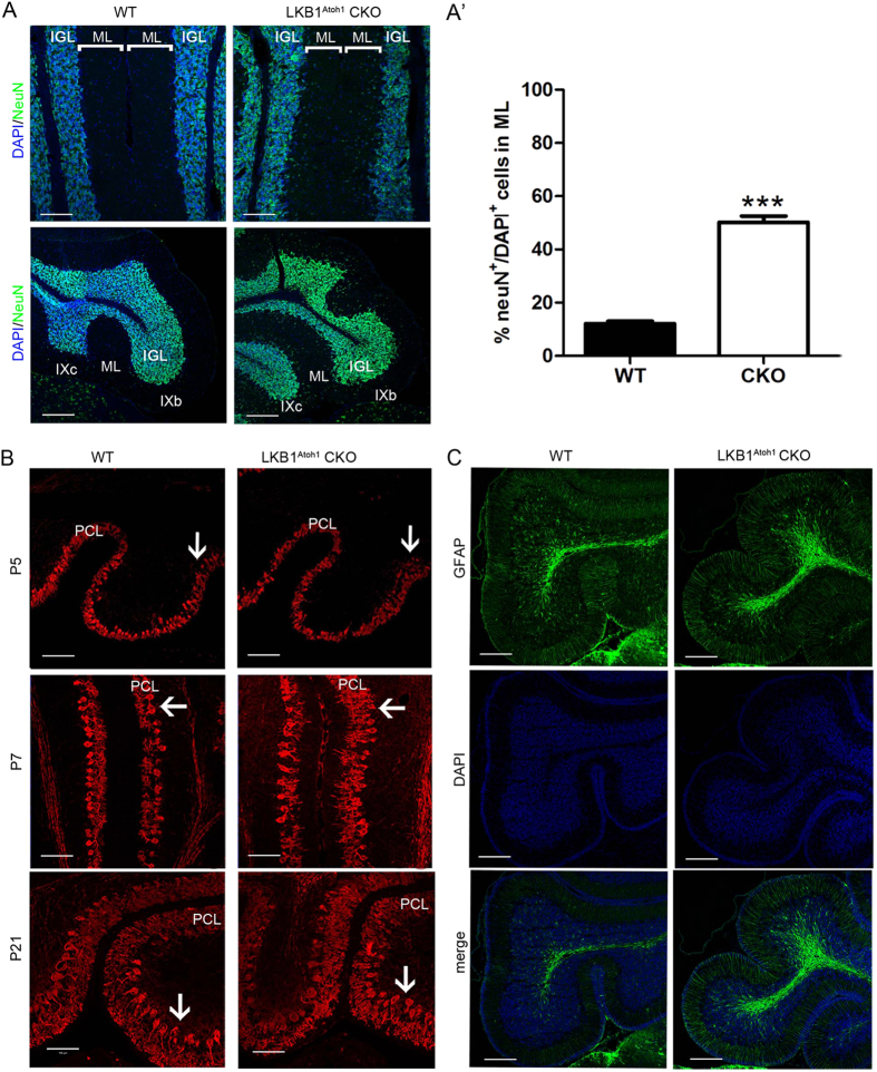
Figure 4. Abnormal granule cells and Purkinje cells in the cerebellum of LKB1Atoh1 CKO mice.
(A) The cerebella of P30 wild-type and LKB1Atoh1 CKO mice were stained with NeuN, a GC marker. The LKB1Atoh1 CKO cerebellum showed an abnormal accumulation of GCs in the ML compared with the wild-type cerebellum. Green, NeuN; Blue, DAPI. Scale bars: 100 μm. (A’) Quantification analysis of NeuN+ cells confirmed an abnormal accumulation of GCs in the ML of mutant cerebellum compared to the wild-type cerebellum (P = 2 × 10 −6). The error bars indicate the SEM. *P < 0.05; **P < 0.01; ***P < 0.001 compared to the WT by Student’s t-test; n = 4 for each group. (B) Aberrant PC development in LKB1Atoh1 CKO mice. CalbindinD28K immunostaining, a marker of Purkinje cells, showed that PCs in the P5 mutant cerebellum were clearly accumulated (arrows) in some parts of the mutant cerebellum compared to the wild-type cerebellum. At P7, the PCs were accumulated (arrows) and showed scant dendritic arborisation (arrows) in the LKB1Atoh1 CKO cerebellum, while the PCs of control cerebellum were present in a single layer. At P21, in contrast to the PCs of the wild-type cerebellum, some of the PCs in the mutant cerebellum were accumulated (arrows) in more than one layer and were disorganised (arrows). Red, CalbindinD28K; Blue, DAPI. Scale bar: 100 μm. (C) Cerebellar section of the P30 wild-type and LKB1Atoh1 CKO mice were stained using the anti-GFAP antibody, a marker of Bergmann glia. No discernible developmental defects were observed in the BGs of the mutant cerebellum compared to the controls. Green, GFAP; Blue, DAPI. Scale bars, 100 μm.

